Multiphoton fluorescence microscopy, in which a fluorophore absorbs more than one photon and emits light at a shorter wavelength than the excitation source, can be used to create 3D tissue images at depths of 1 mm or more. Recently, studies have shown that in vivo two- and three-photon microscopy can image neurons and vascular structures beyond the cortex in the brains of mice.

Imaging the vasculature requires intravenous injection of contrast agents that absorb strongly in the near-infrared and have prolonged blood circulation times. Traditionally, organic dyes are used, despite their poor photostability and low quantum yields. Quantum dots represent a brighter, more stable class of contrast agent, but they introduce substantial toxicity concerns.
Instead, a team headed up at the University of Texas at Austin propose the use of highly fluorescent semiconducting polymer dots (pdots). The 10–100 nm pdots are brighter than traditional fluorophores, have intrinsically low cytotoxicity and can be functionalized for molecular targeting (Biomed. Opt. Express 10.1364/BOE.10.000584).
“We were searching for bright, photostable contrast agents with biocompatible features for in vivo studies,” explains first author Ahmed Hassan. “Moreover, we’re limited to a finite set of excitation wavelengths by the availability of specific laser sources and the absorption and scattering properties of complex, heterogeneous tissue environments. We found that polymer dots fulfilled our requirements and suspected they might be particularly useful for multiphoton microscopy.”
Comparing contrasts
For deep in vivo imaging, three-photon microscopy offers advantages over two-photon imaging, such as suppression of out-of-focus background fluorescence, reduced scattering and improved signal-to-background ratio (SBR). So Hassan and colleagues first identified which excitation wavelengths produce two- versus three-photon absorption in pdots. They examined three pdots — poly(phenylene vinylene)-based CNPPV and fluorene-based PFBT and PFPV — excited by 790–850 nm, 1060 nm, and 1200–1350 nm laser light.
At 800 nm, all three pdots exhibited two-photon fluorescence. For CNPPV and PFBT, this persisted out to 1060 nm, while PFPV began to demonstrate three-photon excitation at this wavelength. Pure three-photon fluorescence was strongest at 1300 nm for CNPPV, 1350 nm for PFBT and 1325 nm for PFPV.
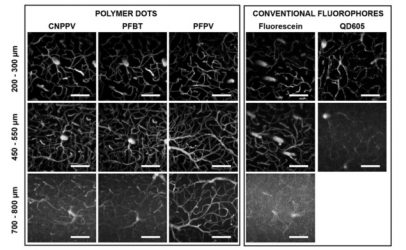
AdvertisementTo evaluate the use of pdots for deep in vivovascular imaging, the researchers injected C57 mice with organic dye (dextran-conjugated fluorescein), quantum dots (QD605), or pdots. They imaged the animals’ vasculature via an optical cranial window, using line scanning and 800 nm excitation from a Ti:sapphire laser.
Comparing 100 μm-thick maximum intensity projections at equivalent cortical depths, the pdot images were brighter than either the fluorescein or quantum dot images. PFPV produced the largest SBR, followed by PFBT then CNPPV. Relative to QD605 and fluorescein, pdot SBR was larger throughout the entire depth range.
The authors note that the mouse injected with inorganic quantum dots did not survive. Conversely, mice repeatedly injected with pdots over several months showed no signs of cytotoxicity or deleterious effects, supporting claims of pdot biocompatibility.
Imaging deeper
Pdots have a wide absorption spectrum that makes them compatible with a number of excitation sources, including longer wavelength lasers that penetrate deeper into tissue. The researchers used 1225 nm excitation (Ti:sapphire laser light shifted by an optical parametric amplifier) to image CNPPV-labelled vasculature.
Excitation at 1225 nm, where CNPPV exhibits a combination of two- and three-photon processes, attained an imaging depth of 1300 µm, compared with 850 µm for 800 nm excitation. The SBR of images collected at 1225 nm greatly exceeded those recorded at 800 nm, for all depths, producing a much higher quality 3D volume.
The team also used a custom-built 1060 nm ytterbium-fibre laser to image PFPV-labelled mouse vasculature. “Fibre lasers are low-cost, high power and available at longer excitation wavelengths, three key features that we believe are necessary for their mass adoption in academic and industry settings,” says Hassan.
While excitation at 1060 nm did not significantly extend the penetration depth, it substantially improved image contrast and increased SBR by about 3.5 times over 800 nm excitation, particularly at depths beyond 600 µm.
The researchers attribute this to three factors: the higher average power of the fibre laser relative to the Ti:sapphire source; the reduced number of photons lost to scattering and absorption at 1060 nm versus 800 nm; and the fact that PFPV exhibits a partial three-photon fluorescence at 1060 nm versus a two-photon excitation signature at 800 nm.
“Overall, pdots present an exciting new approach to multiphoton in vivo imaging,” the authors conclude. “With brighter, biocompatible probes, researchers will be able to resolve vascular architecture in living organisms with improved clarity and depth, enabling critical insights into fundamental biological problems.”
Hassan notes, however, that much work remains before pdots and multiphoton microscopy could be used clinically. “Careful toxicity studies would need to be conducted, and a clearing mechanism for polymer dots has yet to be identified in vivo. In addition, the penetration depth would be limited to shallow cortical layers, meaning endoscopic multiphoton technology would need to be advanced quite a bit for this to be useful in clinical brain imaging,” he tells Physics World.
Source: https://physicsworld.com/a/polymer-dots-image-deep-into-the-brain/
